Physical Description
|
|
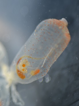
Individual E.diaphanis zooid found on Heron Island, September, 2013. Pigmentation,stomach, intestine and gametes can be seen. |
Ecteinascidia diaphanis colonies are made up of upright zooids attached to each other by short posterior stalk connecting them to common basal stolons. These basal stolons attach to the substrate grounding the animal. Each zooid can reach up to 1cm in height and 0.5cm in diameter. The tunic is very thin and transparent allowing the internal structures to be seen. In animals collected from Heron Island and surrounding reefs it is common to find pink to orange pigment spots within the body wall. These pigment spots are not found in animals living in other areas; however red bands have been noted in other populations. The reasons for these morphological differences remains unclear and even more mysteriously neither pigment spots nor bands are found in preserved specimens (Kott, 1992). Both the inhalant and exhalant siphons are located on the upper surface of the zooid, these siphons are able to open and close allowing for control of the water flow into the pharynx. When open these siphons can be easily recognised by their noticeable, cylindrical protrusion from the body (Kott, 1992).
|