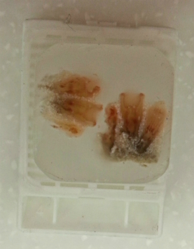
 Internal Anatomy Internal Anatomy 
 The mouth and pharynx opening are elongated slits (Ruppert et.al., 2004). Inside the pharynx has one siphonoglyph with paired ciliated grooves. The siphonoglyphs is responsible for polyp inflation by pumping water in through an inward ciliary movement. The coelenteron partitioned by complete and incomplete unpaired septa. Septal arrangements are unique, new septa only form on either side of the ventral septal pair. In general, each pair of septa include one large septum (macroseptum, extending to the actinopharynx) and a small septum (microsrptum). However, dorsal directives and ventral directives are an exception, which are both microsepta and are both macrosepta respectively. This is called the brachynemous . (Ruppert et.al., 2004)
Tissues and body compartments: Body wall has three tissue layers, epidermis (ectoderm), gastrodermis (endoderm) which lines the gastric cavity and joins the epidermis at the mouth and in between the two epithelia is a gelatinous extracellular matrix named mesoglea. Thick mesoglea with solenia that open into coelenteron and maybe column surface; solenia circulatory channels for thick body wall. (Ruppert et.al., 2004)
Gas Exchange & Excretion: The ciliary flow of fluid over the gastrodermis and epidermis helps gas exchange and the release of ammonia which happen by diffusion over tentacles and the rest of the body.
Musculature: Muscles are arranged in sheets of epidermal or gastrodermal epitheliomucuslar cells. Mesogleal sphincter muscular closes over the retracted oral disc and tentacles. Tentacles have longitudinal epidermal musculature whereas oral disc contain radial epidermal musculature. All the other muscles are gastrodermal with c
ircular muscle in the column wall, longitudinal and radial septal muscles and a circular musculature around the pharynx. The longitudinal septal muscles are call the retractors. The retractors muscles are responsible for in the retraction and invagination of the oral disc and tentacles. The contraction of these muscles can shorten the column and bow the oral disc and tentacles inward, then pull it into the column. Once invaginated, the purse-string-like sphincter muscle constricts the oral disc margin. (Ruppert et.al., 2004)
(Fig 1. Cross section of column and pharynx from Ruppert et.al., 2004)
|