 Cell Biology Cell Biology
Introduction
A unique characteristic of the phylum Cnidaria are their cnidocytes that contain a cnida. Cnidae are one of the defining morphological characteristics that are the most appropriate for higher level taxonomy and to discriminate species (Ryland & Lancaster,2004). One category of cnidae are the nematocysts. Thick-walled nematocytes occur in all group of cnidarians, they generally have spines or barbs on the surface of the everted tubule (Ruppert et.al., 2004). The wall of nematocyst is thickened and stiffed by a layer of collagen. The nematocysts degenerates and is replaced by a new one after discharging the nematocyst, so there is a high energetic cost to fire each tubule (Ruppert et.al., 2004). There are at least 7 types of nematocysts are know for Zoanthidea (Ryland & Lancaster,2004), this study aim to find and identify the type of nematocytes found in P.caesia and discuss the use of the nematocytes.
 Method Method
P.caesia were collected from from the reef flat and outer slope of reef crest on the southside of Heron Island (23º27'S, 151º57'E), removed using a spatula and relaxed by relaxant agent. Then fixed with 0.02% of MS222 Tricaine in 100ml of seawater to prevent damage of tissue structure. Fixed specimens were then place in 70% ethanol and trimmed into “minimal” tissue. Small sample were placed in basket with label of sagittal section and sent off for sectioning. The sections were stained with haematoxylin and eosin before analysis under microscope.
Result & Discussion
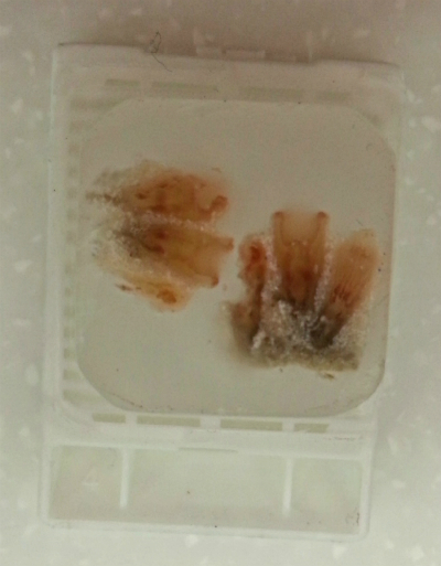
Nematocysts were found on the tentacles of P.caesia. Nematocysts tend to locate on the edge of the tentacles. The Nematocysts was large and oblong-oval capsules and have two different tubule. First type is called Holotrich I, tubule was very wide with large spines organise in a tripe helix when discharged and it was very loose and obliquely coiled (Ryland & Lancaster,2004). The second one is called atrich, which has a thinner tubule and lack of spines, before discharge the tubule was coiled in a spring like capsule (Ryland & Lancaster,2004).

Nematocysts were found in abundance on the tentacles, showing that they play a central role in capture prey, defense against other organism and aggression against competitors. The nematocysts present on the tentacles are important as they are used in the capture and paralyze active prey. When the prey is captured by the tentacles, nematocysts may sting the prey and inject toxins to paralyze prey. Nematocysts are used for feeding, defense and aggression, all of which are important for fitness which enhance their survivorship.
.jpg) (Fig. 2 Nematocytes found in specimens)
(Fig. 2 Nematocytes found in specimens) |