Physical Description
General Echinoid Description
Sea urchins display pentamerous symmetry (Ruppert et al. 2004). Their body is formed by skeletal elements (interlocking plates),and is referred to as a test (Nichols 1967). The test is composed of alternating double columns of ambulacral and interambulacral plates (Nichols1967). Their test is covered with five double rows of podia (Leddy & Johnson 2000; Ruppert et al. 2004), which are connected through pores in the ambulacral plates (Nichols 1967; Ruppert et al. 2004). This feature is unique to echinoids and their interambulacral areas lack tube feet (Ruppert et al. 2004). Their characteristic Aristotle’s lantern, which consists of five teeth, is found on the oral surface of the animal as it functions in feeding (Nichols 1967). Sea urchins have spines arranged all over their body, these vary in size and colour between species (Ruppert et al. 2004). Radiating from the sea urchin test are tube feet (podia), which have a variety of functions including feeding, locomotion, sensory perception and adhesion (Flammang et al. 2005; Leddy & Johnson 2000; Nichols 1971; Smith 1979) and are an extension of the water-vascular system (WVS) (Ruppert etal. 2004).
Distinguishing Features of Temnopleurus alexandri (as described by Miskelly 1968)
T. alexandri displays a variety of test and spine colours. The test is generally light shades of colours such as green, purple and white. The colours of the spines vary from the base of the spine at the test to the white tip; the combinations of these can be green and purple, purple and white, white and maroon and green and brown. The test can grow as big as 86mm in diameter and be described as conical in shape. The spines are equal in length around the whole test with the maximum length being 12mm. The spines at the oral axis of the test are spatulate in shape, whereas the aboral spines taper to a point. The tube feet of T. alexandri were observed to extend more than three times the length of the spines.
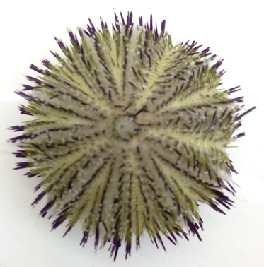 |
View of aboral surface of Temnopleurus alexandri
Photo: Monique Parisi
|
Spines (Information summarised from Ruppert et al. 2004)
Sea urchins generally have two types of spines distributed over the body; these are known as primary spines and secondary spines, with primary spines being longer than secondary spines. The spines are connected to the test by a ball-and-socket joint, which allows them to move in a variety of directions. This movement is enabled by the presence of outer muscular sheaths and inner collagen fibres, which also allows them to lock the spine in place. Spines can vary in shape, length and colour between species, but they are usually cylindrical and taper to a point. The spines have regenerative capabilities and play a key role in defense and wave resistance. They also function in locomotion.
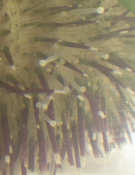 |
Close up of spines (purple) and tube feet (transparent) of Temnopleurus alexandri
Photo: Monique Parisi
|
Pedicellariae (Information summarised from Ruppert et al. 2004)
There are a range of pedicellariae with different functions present all over the body of sea urchins. They are generally a group three jaws mounted on a stalk, which are controlled by muscle contraction and stimulated by physical contact, either on the inside or the outside of the jaws. Pedicellariae are involved in defense, self-grooming and breaking up of food particles. They may also function in chemosensory reception as they respond to chemical stimuli.
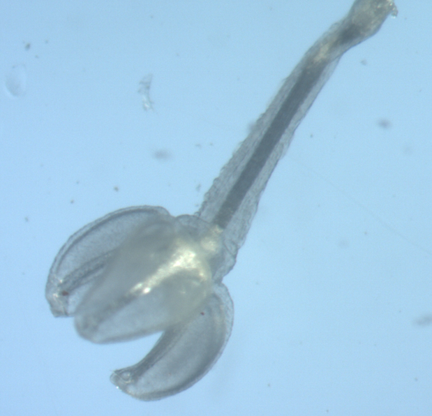 |
Temnopleurus alexandri aboral pedicellaria
Photo: Monique Parisi
|
|