Life History & Behaviour
Background information
Feeding
Like most other solitary ascidian, the yellow sea squirts are suspension feeders.
They feed by absorbing water through their oral siphon with the help of cilia which are tiny hair-like appendages found on the edge of the gill slit. Cilia create a current which pump water into the pharynx through a mucus net, trapping the suspended particles. This mucus net is produced by the endostyle and located inside the branchial basket of the ascidian (See anatomy). The water will then exit the ascidian via the atrial siphon (Peterson 2007, Kott 1985).
Respiration
Similarly, the water is also taken to the gill slits where respiration takes place (Ruppert et al. 2004).
Reproduction &life cycle
Phallusia julinea is a species of solitary ascidian found on the Heron Island reef and is hermaphrodite as all ascidians. Solitary ascidians are oviparous having both an egg sac and sperm duct. Fertilization occur in seawater where the eggs and sperm are released through the atrial siphon (Kott 1985).
Ascidian larvae are free-swimming, they move with their post-anal tail, just like any other chordates. The larval phase, which can last from minutes to hours, correspond to the time needed for the larvae to reach competency and find a suitable habitat to settle and go through metamorphosis. Once settled, the ascidian becomes a sessile organism and does not move for the rest of its life (Jeffery 1997, Roberts et al. 2007).
Experiments
The time it takes for the eggs to be fertilized and the larvae to develop usually differ according to the species, as does the competence of the larvae (Davidson & Swalla 2002). The following experiment was designed to watch and record every stage of the Phallusia julinea development as well as how much time the larvae need to settle.
Methodology
Egg fecundation and development
Three Phallusia julinea specimens were collected in the field, on the Heron Island reef crest on the 17thSeptember 2012 and brought back to the Heron Island Research Station.
Two of the three specimens were dissected in order to have access to the egg sacs and sperm ducts. Both the egg sacs and sperm ducts were pierced using a scalpel and the eggs and sperm from both specimens were aspired using a glass pipette. They were then transferred to a petri dish filled with seawater, and the water was stirred so the gametes from the two individuals could mix properly and maximize the chances of fertilization.
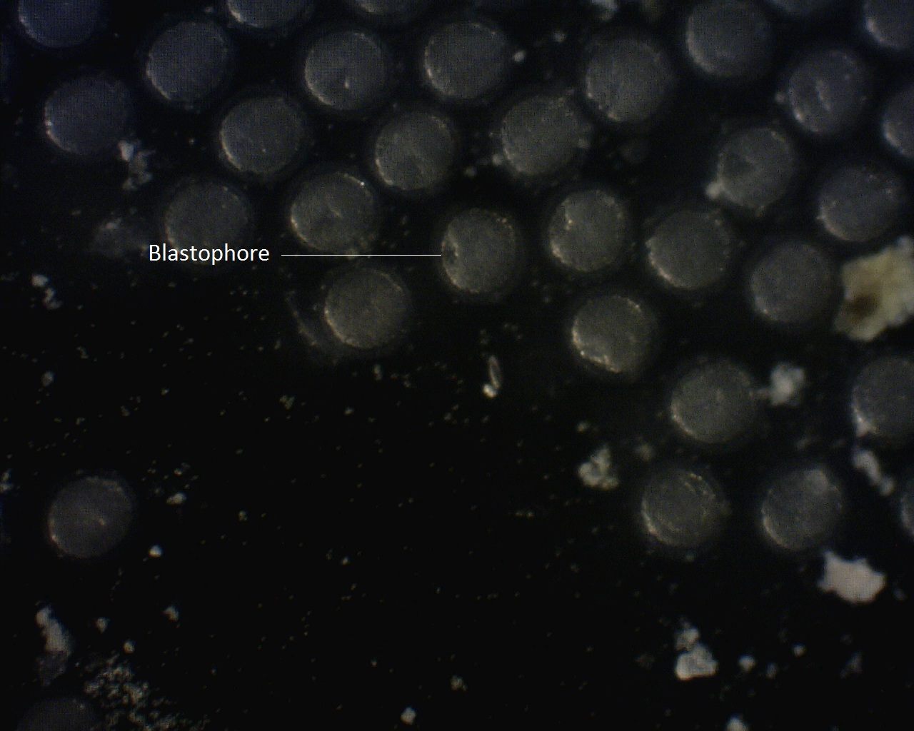
The petri dish was then placed under a dissecting microscope and monitored to spot any signs of fertilization, and then any stages of cell division possible, as well as gastrulation, creation of the blastophore and apparition of the tail and the eyes. Pictures were taken using a Dino-Lite camera placed on one of the eye socket of the microscope.
Larvae settlement
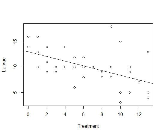
Several six-well dishes were filled with 5 mL of seawater and each column of three wells given a number, starting with zero and increasing by one every column. Twenty fertilized eggs were placed in each dish. As soon as the eggs hatched, the second experiment started. Every hour, small coral rubble was added to each dish of a column, starting with column 1 at time 0. Column 0 received no rocks and acted as a control. After about 17 hours, the number of unsettled larvae was counted in each dish in order to determine the total number of larvae that settled. A linear regression was then performed to test the significance of the change in settled larvae in each column.
Some of the settled larvae were then taken back to the University of Queensland to determine how much time they take to go through metamorphosis and change into fully functional ascidians.
Results
Egg fecundation and development
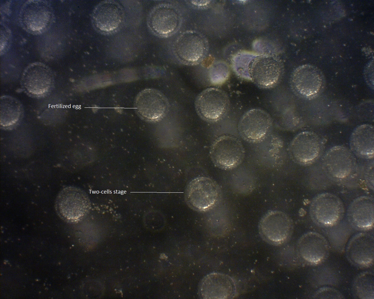
After fecundation,the eggs were fertilized rapidly but dueto the transparency of the gametes, itwas impossible to determine an exacttime. Table 1 presents different stages inthe development of the eggs, and thetime taken from fecundation to get to thisstage.
Table 1: Different stages of the ascidian development andthe time taken to reach it after fecundation
|
Stage
|
Time (Hours:Minutes)
|
|
2 cells stage
|
0:45
|
|
4 cells stage
|
1:20
|
|
8 cells stage
|
1:40
|
|
16 cells stage
|
1:55
|
|
32 cells stage
|
2:15
|
|
Blastophore visible
|
4:10
|
|
Tail bud starts to appear
|
8:00
|
|
Eyes start to appear
|
12:15
|
|
First hatchling
|
14:55
|
Larvae Settlement
No larvae in the control dishes settled, but there was asignificant decrease in free living larvae as the time at which the rubble wasput in the dishes increased (p =0.045)(Fig. 1). 90% of the larvae died on the way to the University of Queensland. Amongst the few survivors, one larva managed to complete itsmetamorphosis seven days after fecundation. The ascidian was still a juvenile,with no pigmentation and just a few millimetres big, but both siphons werevisible and fully functional. Because of the absence of pigmentation, the internal anatomy of the sea squirt could be directly observed.

Figure 1. Influence of time on larvae settlement.
Discussion
The fertilized eggs took about fifteen hour to hatch intolarvae, which correspond to Jeffery’s (1997) estimations of the amount of time it should have taken them to hatch. The time it took for the larvae to become competent could not be calculated exactly for it was impossible to determine when exactly the larvae settled on the coral rubbles. Larvae in contact with coral rubbles before they become competent could be less likely to settle on the rubbles because they got used to them. The fact that more larvae seemed to settle in wells where the rubbles were introduced later suggests a rather high period of time before they become competent. These results need to be confirmed with the help of a more thorough experiment.

Photo of fertilized eggs and first cell division

Photo of fertilized eggs and second cell division

Creation of the blastophore

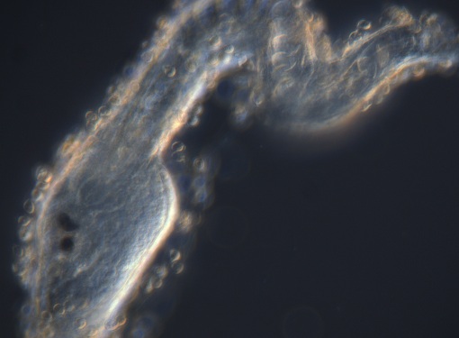
Ascidian Larvae just after hatching
Did you know? Ascidians can absorb their own body volume of water every second. |