Internal Anatomy
Digestion (Foster 2006):
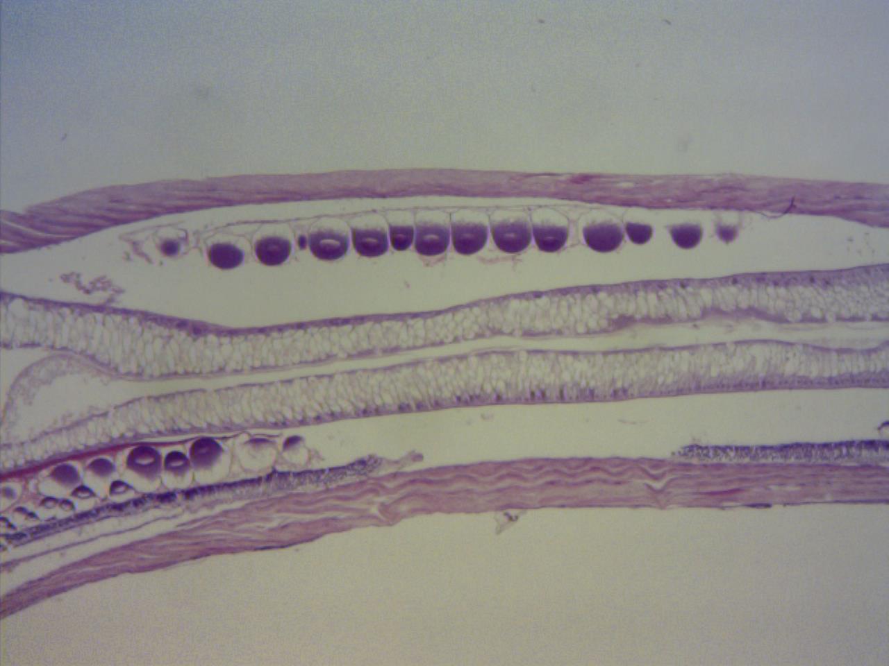
Chaetognaths contain a simple intestine that runs the length
of the trunk (fig 1), and connects to the rectum which is located at the trunk-tail
boundary. The expulsion of faeces is controlled by a rectal sphincter that is
composed of circular muscles.
Nervous system:
Chaetognaths possess a well developed, ganglionated nervous
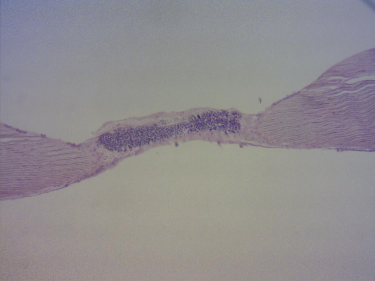
system which is made up of six ganglia on the head and a large ventral trunk
ganglion (fig 2) (Ruppert et al 2004, Foster 2006). The largest ganglion is the ventral
ganglion, which is located in the trunk of chaetognaths, and acts to innervate
the muscles of the trunk and coordinate swimming.
Sensory system (Foster 2006):
The sensory organs include eyes, ciliary tufts, and the
ciliary loop (corona). Chaetognaths exhibit phototaxis, and it is therefore
believed that it is the eyes that play a major role in this behaviour. There are
lots of ciliary tufts located along the head, trunk and tail of chaetognaths. The
main role of these ciliary tufts is to detect prey in the water column. It is
not fully understood what the exact purpose of the ciliary loop is, however
multiple roles have been suggested, and these include: excretion of metabolic
wastes, chemoreception or to guide sperm to the gonopores.
Muscular system (Foster 2006):
Depending on the species in question, there are approximately
16 head muscles which assist in manoeuvring the grasping spines, teeth and mouth
during feeding.

Figure 1: Dissection microscope view of the digestive tract of a stained chaetognath.

Figure 2: Dissection microscope view of the ventral ganglion of a stained chaetognath. |