External Morphology
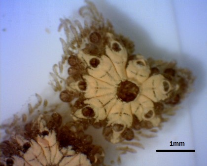
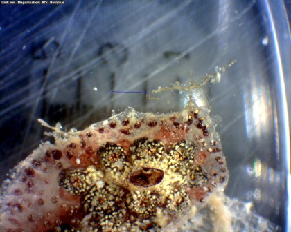
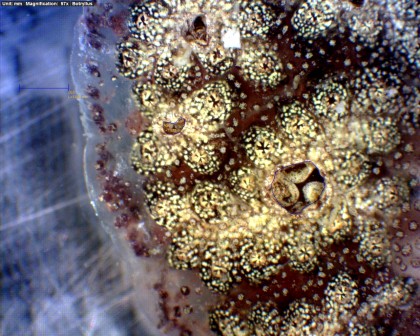
B. schlosseri is characterised by star-shaped systems of zooids embedded in a common tunic. The tunic is made primarily of tunicin, a polysaccharide, which gives the colony a fleshy texture (Carver et al. 2006). Zooids share a vascular system made of blood vessels that terminate in pigmented ampullae on the periphery of the colony. Individual zooids have their own buccal siphons but share an atrial siphon with other zooids in a system. Colonies of botryllus may take on a variety of colour morphs. The expression of these colours is controlled by alleles at a few loci (Morris et al. 1980). 3 colour morphs observed at Heron Island are shown below.
  
Figure 3: 3 distinct colour morphs of B. schlosseri encontered on Heron Island, September 2011. Scale bars represent 1mm
Heron Island Research Project
On Heron Island, the number of zooids per system within a single colony varied from 4 to 13. The mechanism whereby primary buds migrate together during takeover to form a common atrial siphon is poorly understood (Grosberg et al. 1987). It is possible that individual zooids each contribute a determinate amount to the construction of the apical aperture. If this is the case, we would expect to see a linear relationship between the cross sectional area of the apical aperture and the number of zooids that share that aperture. A simple experiment was conducted to determine whether the number of zooids present in a system was related to the cross sectional area of the atrial siphon in colonies of Botryllus schlosseri.
Methods
Coral fragments with encrusting Botryllus schlosseri colonies of a particular colour morph were collected from the reef crest of northern Heron Island during low tide over a period of 3 days in September 2011. Fragments were kept in storage tanks with ambient conditions at the Heron Island research station. Intact colonies were gently removed from the coral fragments and transferred to petri dishes. To ensure a that atrial apertures remained completely open for the taking of measurements, colonies were anaesthetized in a 0.02% solution of MS222. Anaesthetized colonies were viewed under a light microscope and photos of each system in the colony were take with a Dino-Lite microscope eyepiece camera. From the images, the number of zooids per system was counted and the cross-sectional area of the atrial aperture was estimated using Dino-Lite software.
 
Figure 4: Photographs of anaethetized zooid systems of B. schlosseri taken with Dino-Lite microscope eyepiece camera
Only functional zooids with open buccal siphons were counted to ensure a standardised measure for zooid number. Linear regression analysis was performed to determine whether a significant relationship exists between cross sectional area and the number of zooids per stellate system.
Results

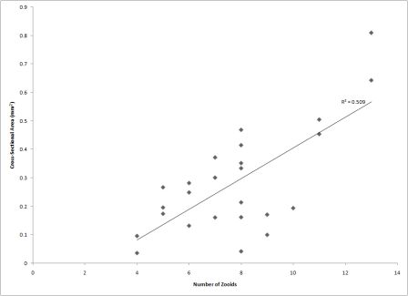
Figure 5: relationship between number of zooids in a system of B. schlosseri and the cross-sectional area of that atrial aperture
The cross-sectional area was estimated for 25 systems of zooids from 5 colonies of B. schlosseri. Cross-sectional area was not significantly different between colonies (p = 0.1431). The linear regression analysis showed an insignificant positive correlation between the number of functional zooids in a system and the cross-sectional area of the apical aperture (R² = 0.509; p = 0.15). From this study, we can conclude that the size of the apical aperture for a single system is indeterminate of the number of zooids.
|