TUBE FEET
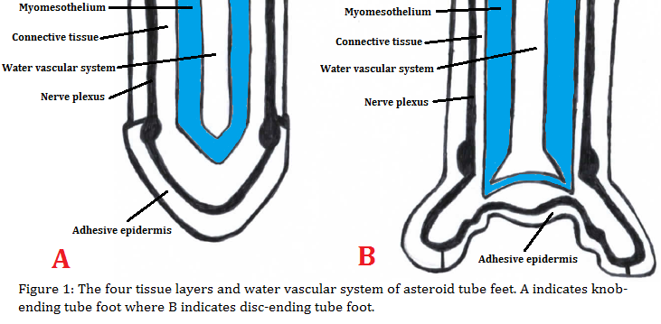
The podia, also known as tube feet, are highly specialised structures that aid echinoderms in locomotion, fixation, burrowing as well as adhering to substrates (Santos et al. 2005). The anatomy of tube feet consists of a basal extensible cylinder, or stem, which expands to form an apical flattened disc or ‘sucker’ (Santos et al. 2005) (Figure 1). Each tube foot consists of four tissue layers that are specialised for adhesion and sensory perception : the inner myomesothelium, a connective tissue layer, the nerve plexus and the outer epidermis that is covered by the cuticle (Santos et al. 2005)(Figure 1).
As tube feet may perform different functions, the shape or form of tube feet of various echinoderms often correspond to a particular function. Asteroid tube feet may be identified by two existing forms – ‘knob-ending’ podia that have pointed tips and ‘disc-ending’ podia that have flattened tips (Santos et al. 2005)(Figure 1). The tube feet of Gomophia watsoni are that of the latter, having very distinct, flattened suckers at the tip of the podia. The variation in tube feet shape and form may therefore correspond to the substrate type or environment they are likely to inhabit, such as soft or hard substratum. Given that G. watsoni inhabits rough surfaces such as rocks, it is therefore fitting that it bears disc-like suckers.

Adhesion
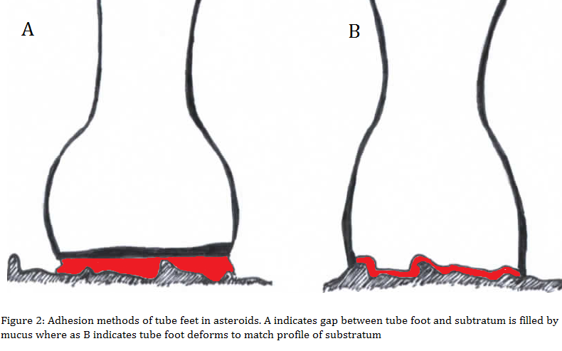
Like most echinoderms, G. watsoni relies upon the adhesive capabilities of tube feet to adapt to environmental conditions and constraints such as rough substrates and harsh tidal conditions. The tube feet act as external appendages of the water vascular system that are able to adhere strongly to various surfaces by means of mechanical suctioning, chemical interactions and the secretion of adhesive mucus from the tip of the tube feet (Harrison & Philpott 1966; Thomas & Hermans 1985; Vickery & McClintock 2000). Approximately just over half of the adhesive strength of asteroids is due to the mechanical suction of tube feet, while the rest is attributed to adhesive secretions (Engster & Brown 1972). The flattened disc at the end of the stem continually attaches and detaches from various surfaces by means of secreting adhesive and then de-adhesive mucus produced by specialised cells (Santos et al. 2005). These specialised cells are located in the epithelium of the tube feet suckers (Engster & Brown 1972). Asteroids may then attach to the substrate by either maintaining a flat position and filling the gap between the base of the sea star and the substratum with mucus, or by physically moulding to match the profile of the surface (Santos et al. 2005)(Figure 2). The functionality of asteroid podia are integ ral components of their anatomy, without which would be unable to move, attach, capture or handle prey. It may therefore be inferred that the morphology of tube feet of G. watsoni and other asteroids may be an adaptation to occupy certain niches, habitats and substratum in which echinoderms may be found. ral components of their anatomy, without which would be unable to move, attach, capture or handle prey. It may therefore be inferred that the morphology of tube feet of G. watsoni and other asteroids may be an adaptation to occupy certain niches, habitats and substratum in which echinoderms may be found.
Locomotion
G. watsoni relies heavily upon the functionality of tube feet for locomotion and movement. Movement is generated by extension-attachment, force-generation and detachment-retraction (Ruppert et al. 2004). As the ampulla muscles contract, the valve in the lateral canal closes which fills the tube foot with water. As the foot comes into contact with a surface, the apical sucker then adheres via suction and mucus secretion. To detach, muscles are retracted causing fluid to be displaced within the ampulla, with the assistance of de-adhesive mucus. Such adhesion and de-adhesion by individual tube feet contributes greatly to the movement and locomotion of G. watsoni. Each tube foot ‘steps’ by moving backward and forward while adhering which allows the seastar to coordinate movement in a particular direction. During movement, one or two arms may lead directionally while the others follow suit. This directional movement and strong adherence of G. watsoni effectively allows the species to attach and climb on surfaces of varying roughness and angles. This evidently contributes greatly to its ability to inhabit small and irregular surfaces such as rocks and coral.
Tube Feet Microscopic Analysis
To properly examine and analyse the anatomy of G. watsoni, a microscopic analysis was undertaken to better understand the morphology and physiology of its tube feet. A G. watsoni specimen was obtained from the field in the reef crest on Heron Island, Australia. When handling the specimen, tube feet were generally unseen as they were contracted. To encourage the specimen to relax and reveal its tube feet, the sea star was slowly exposed to increasing quantities of magnesium chloride until tube feet started to emerge. When fully relaxed, the specimen fully extended all its tube feet. Observations showed that the tube feet of G. watsoni were knob-ending podia with distinct flattened suckers with fairly long stems in comparison with arm diameter. One or two tube feet were carefully removed for the purposes of DNA barcoding.
To further investigate the internal anatomy of G. watsoni, a dissection was conducted. However this was fairly unsuccessful given the very small size of the species it’s very hard outer cuticle that made delicate incisions difficult. However, various parts of the water vascular system and internal appendages were observed. Finally, an arm was carefully amputated for the purpose of sectioning and staining to gain a better understanding of the internal anatomy of G. watsoni. However, sectioning was unsuccessful due to the inability of the fixative to penetrate the tissue while the endoskeleton did not soften.

|