Overview
Brief Summary
Common Names
Comprehensive Description
Biogeographical Distribution
Physical Description and Morpholgy
Features of a Planaxid Shell
Size
Shell Morphology
External Anatomy
Internal Anatomy
Apomorphy
Evolution & Systematics
Fossil History
Phylogenetics
Ecology
Local Distribution and Habitats
Micro-habitats and Associations
Life History & Behaviour
Larval Development
Reproductive Behaviour
Locomotion and Foraging Behaviour
Predator Avoidance and Escape Behaviour
Conservation
Trends and Threats
References & More Information
Bibliographies
Search the Web | Internal Anatomy
|
The Columellar Muscle
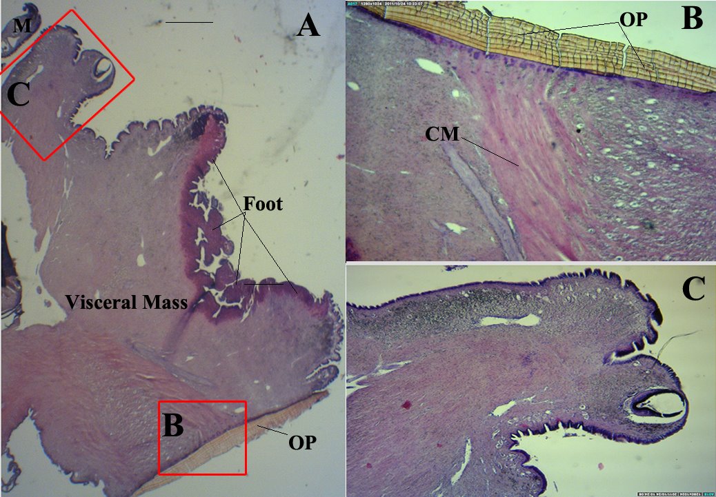
The Columellar muscle is the largest, most prominent and most important muscle in the anatomy of a marine gastropod (Ruppert, Fox & Barnes 2004). The muscle is responsible for holding the soft body parts of the animal in place in the shell and withdrawing the animal into its shell when required (i.e. Protection from predators and drying out) (Ruppert, Fox & Barnes 2004). The Columellar muscle originates and attached strongly to the columella of the shell and inserts on the operculum in the foot (Ruppert, Fox & Barnes 2004).
|
| |
 |
|
| |
Longitudinal Cross section (LS) of P. sulcatus head-foot visceral mass. (A) Cross section of the head-foot region (10X magnification). Note the large buccal mass filling most of the downward-pointing snout, operculum, mantle and contracted foot. (B) Close up showing attachment of the collumellar muscle to the operculum (100X magnification). (C) Close up of the snout (10X magnification). Note the large buccal cavity in proportion to the snout and presence of a pair of tiny jaws. OP= Operculum, M=Mantle, CM= Collumellar Muscles. Sections are stained with Mayer’s Haematoxylin and Eosin.
|
|
|
Alimentary tract
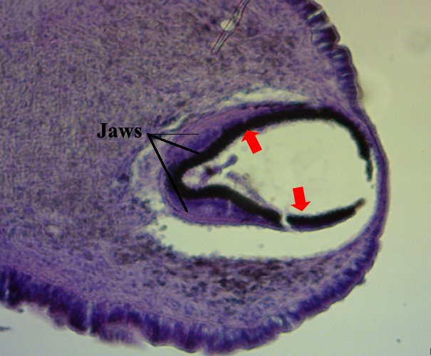
The buccal mass is very large and well developed filling the snout (Houbrick 1987). The mouth is located in the concave ventral portion of the oral hood and appears as a longitudinal slit (Houbrick 1987). There is a pair tiny jaws, made of chitinous scale-liked pieces at the edge, on the dorsal side of the inner lips (Houbrick 1987).
|
 |
|
Longitudinal Cross section (LS) of the snout on P. sulcatus. Note
the presence of a pair of jaws made of chitinous scale-liked pieces
at the edge, on the dorsal side of the inner lips. Arrow points to
the chitinous scale-liked pieces lining the edges of the inner lips.
Sections were stained with Mayer’s Haematoxylin and Eosin.
|
|
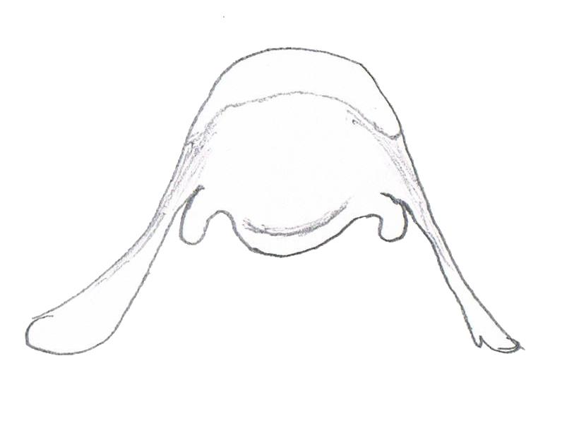
A long, robust radula curves under the buccal mass and dorsally around the nerve ring, terminating in the radula sac, which lies to the right of the nerve ring (Houbrick 1987). The radula ribbon is long and has about 5 rows of teeth/ mm (Houbrick 1987). The rachidian tooth is squarish in shape, with a pentagonal basal plate on which pair of basal lateral cusps, a rounded basal margin and a long thin lateral extension on each side as illustrated below (Houbrick 1987). The cutting edge is convex with a single broad spoon-like cusp (Houbrick 1987).
|
 |
|
Illustration of a rachidian tooth from
P. sulcatus. Modified from Houbrick (1987).
|
|
A pair of minute salivary glands, which appears as coiled tubes, lies together in close proximity spread over the anterior dorsal surface of the esophageal gland and appear separate before emptying into the dorso-lateral sides of the buccal mass (Houbrick 1987).
The esophageal gland is formed by the lateral expansion and thickening of the mid-esophagus (Houbrick 1987). The inner wall of the esophageal gland is folded into many deep longitudinal lamellae (Houbrick 1987). The esophageal gland extends posteriorly nearly the length of the mantle cavity before constricting to form the posterior esophagus (Houbrick 1987).
|
| |
|
|
| |
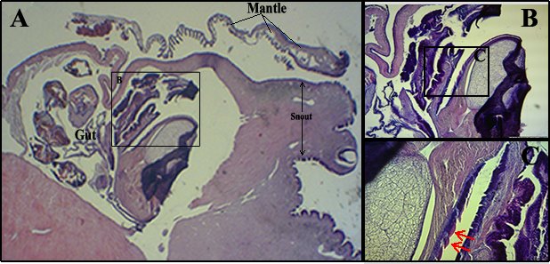
Longitudinal Cross section (LS) of P. sulcatus head-foot visceral mass. (A) Cross section of the head-foot region (10X magnification). (B) Close up of the esophageal gland within the gut region in the visceral mass (100X magnification). (C) Inner wall of the esophageal gland (400X magnification). Epithelial cells lining the esophageal gland stain deep purple with hemotoylin. The inner wall of the esophageal gland is folded into many deep longitudinal lamellae. Arrows point to the longitudinal lamellae lining the inner wall. Sections were stained with Mayer’s Haematoxylin and Eosin.
|
|
|
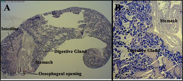
P. sulcatus has a very large stomach (Houbrick 1987). The stomach has a wide, highly muscular and highly mobile, posteriorly bilobed T-shaped raised pad to separate particulate matter entering the stomach from the oesophagus (Houbrick 1987). Two typhlosoles (inner fold of the intestine) are located in the anterior portion of the stomach and is weakly ciliated (Houbrick 1987). A single opening to the digestive gland is close to the esophageal opening and lies to the left base of the bilobed pad (Houbrick 1987).
|
| |
 |
|
| |
Longitudinal Cross section (LS) of the posterior visceral mass of P. sulcatus. (A) Overview of the posterior visceral mass (10X magnification). Note the position of the esophageal opening, stomach and intestine in this part of the visceral mass forming the U-shape gut. (B) Glandular cells of the Disgestive gland (400X magnification). Sections were stained with Mayer’s Haematoxylin and Eosin.
|
|
|
Nervous system
P. sulcatus have an epiathroid nervous system consisting of large major ganglia (Houbrick 1987). The subesophageal ganglion is attached to the left pleural ganglion and the visceral ganglion lies at the posterior end of the mantle cavity, forming a ganglionic mass to the right of the esophagus and a secondary mass crossing over the oesophagus (Houbrick 1987). The cerebral-pleural connectives are very long (Houbrick 1987). The pedal ganglia are large and each has two swollen extension/strand with external one being more developed than the internal one (Houbrick 1987). The RPG ratio is an index that measures the concentration of the nervous system (Houbrick 1987). A high RPG ratio is indicative of a loose condition that is considered plesiomorphic/ancestral whereas a low ratio indicates a tight and more advance condition (Houbrick 1987). The RPG ratio of P. sulcatus is reported to be 0.57, hence suggesting the presence of a tight and well developed nervous system (Houbrick 1987).
|
|
Reproductive system
The pallial gonoducts of P. sulcatus are tubular extensions of the coelumic gonoducts develop from tissues of the base and right wall of the mantle cavity (Houbrick 1987). The pallial gonoduct is an open, slit tube comprising of two adjacent laminae joined together dorsally (Houbrick 1987). The laminae are consists of loosely organised connective tissues covered by epithelium and contain the reproductive structures formed as secular invaginations of the epithelium (Houbrick 1987).
In males of P. sulcatus, the pallial gonoducts is simply an open tube with a proximal glandular prostate (Houbrick 1987). In females of P. sulcatus, the pallial oviducts have a proximal albumen gland and a median capsule gland and the oviducal groove is moderately glandular along its length (Houbrick 1987). At the distal end of the median lamina, a long sperm gutter extend to the median edge and enters the lamina as a duct that branches into a large proximal spermatophore bursa and a smaller, median seminal receptacle lying beneath the spermatophore bursa (Houbrick 1987). A highly ciliated groove emerges from the distal pallial oviduct and runs down the right side of the foot into the birth pore of the brood pouch (Houbrick 1987). The brood pore is a slit in the center of an elevated, lightly pigmented portion of the neck, beneath the peduncle of the right cephalic tentacle (Houbrick 1987).
The brood pouch is consists of a large, primary chamber subdivided into many secondary, lamellar chambers that fill both sides of the head-foot (Houbrick 1987). In P. sulcatus, embryos are found throughout the entire brood pouch and may comprise several cohorts of different growth stages (Houbrick 1987). The more advance embryos are located in the dorsal portion of the chamber (Houbrick 1987).The development of the embryo and larvae is being further described in the larval development page.
|
|
|