Visual Behaviour
Introduction
Pseudoceros ferrugineus is a species of flatworm, belonging to the phylum Platyhelminthes and class Turbullaries, which is commonly found in tropical and subtropical waters (Newman and Schupp, 2002). Although behavioural studies for this species are limited, primarily due to only sporadic discovery in few places across the world, extensive studies have been conducted about Turbullarian vision.
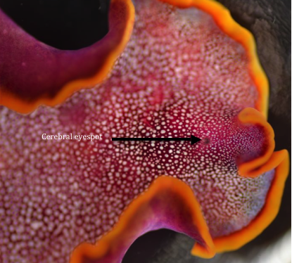
So far it is known that free-living flatworms have photosensitive cells, which are found grouped on the head in a complex arrangement referred to as the cerebral eyespot, which can be seen in figure 1 (Hyman, 1959; Eakin and Brandenbergen, 1981). This simplistic eye is known as a pigment-cup ocelli and the pigmented light sensing organs within the cerebral eye spot are known as ocelli (Sopott-Ehleus, 1991). Each ocelli is made up of a few photosensitive cells as well as a concave cup.
These “cupped shaped” organs project light into a light sensitive cell (microvilli) which is located close to the brain (Sopott-Ehleus, 1991). The cup allows light to enter from three sides, and because of the arrangement of microvilli inside the cup shape cell, light can only come from one direction (Eakin and Brandenbergen, 1981).
The angle of the light, which penetrates the microvilli, then leaves a shadow and because the cup-shaped organ is muscularised it can be rotated to give a three dimensional shape (Hyman, 1959; Eakin and Brandenbergen, 1981; Newman and cannon 1990).
The aim of this study was to test whether or not stimuli in the form of different coloured lights can influence the reactional behaviour of P. ferrugineus.
  
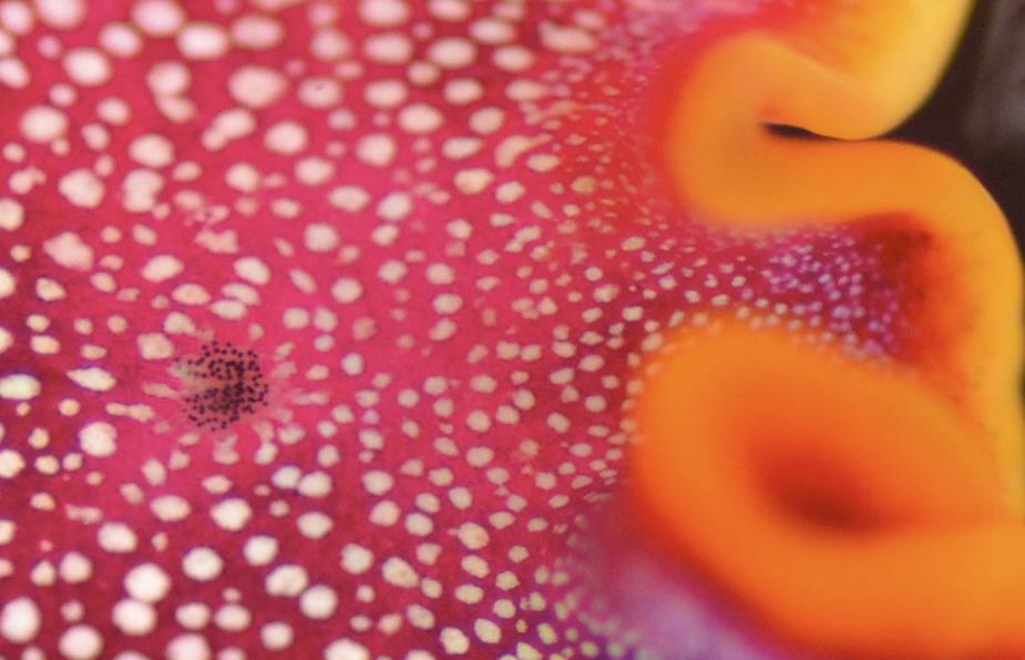
Figure 1: The cerebral eyespot can be seen in this P. ferrugineus as a small cluster of black cells
Method
Three organisms were collected at the southern end of Heron Island at the reef crest, using a plastic container. The organisms were handled with great care as flatworms are notorious for breaking apart. Once these organisms were transported back to the laboratory, they were placed in a glass fish tank, in which they lived for three days, before being released back where they were found. The experiment was in a plastic container, which measured 17cm in length and 7cm in height. This was then spray-painted black around all sides except one, in order to block out all natural light. One end was left clear so that artificial light could shine through. The artificial light emanated from an external microscope light and was placed 1cm away from the container to ensure maximum light penetration. This allowed for a gradient of light to occur with the brightest area being closer to the light and a gradual decrease in light intensity the further away from the primary light source (This set up can be seen in video 1).
Four different parameters were used, namely white light, red light, green light and no light (control). The green and the red light were filters that were obtained from Lee Filters. The green was Oregon Green 488 and the red was Primary red 106. The white light was the external microscope light and the control consisted of blacking out all sides of the container to see if the organism freely moved towards either end without a light source.
The flat worms were set to start in the middle of the container with a 25mm joint covering the animal (As seen in video 1). This ensured that the animal started exactly in the middle. Once the joint was lifted, a stopwatch timed how long it took for the animal to reach the end of the container (either towards or away from the light). The time was recorded and the animal replaced into the holding tank. This was done to each animal, for each trial, for the two primary time periods (day and night). These experiments ran over 2 days which allowed the gathering of data from two days as well as two nights.
Video 1: This video shows how the experiment was set up and the reaction Pseudoceros ferrugineus had towards different types of light. (This video has been sped up by a factor of 2)
Results

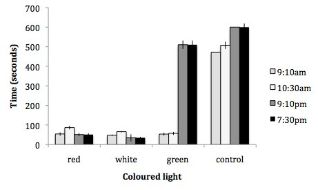
Figure 2: A comparison of the time it took for three individual P. ferrugineus’s to move towards different coloured lights. The different coloured bars represent the different times during the day and night. This graph shows how long it took each individual to move towards the coloured end as well as the individuals which did not move at all. Once the time reached 600 seconds and the animal had not moved either towards or away from the light (or where the light would have been in the case of the control experiment) the animal was thought to have spent a significant amount of time inactive to be disregarded.
It can be seen in figure two that the animals responded the fastest in a positive manner towards the stimuli of both red and white light. The average for the red light during the night and day was 60 seconds with the white being 45 sconds. The control took the longest time for the animal to react with both the 7:30pm and the 9:10pm animals not reacting at all. The green light during the day took an average of 53 seconds and 54 seconds, whilst at night it took over 500 seconds.

Table 1: This table shows the amount of times each individual went towards the light in a percentage.
It can be seen in table one that the red and white light have the most consistent percentage of P. ferrugineus going towards each light respectively. The green light shows that during the morning the organisms go towards the light 100% of the time, whereas later at night the organisms only go towards the light 33% of the time. The control shows that, although 66% of the organisms headed towards where the light would have been during the day time, no organisms moved at all during the night.
Discussion
In figure two the results show that individuals showed a preference towards both the red and white light during both night and day, as well as the green light during the day only. This is evident with the average times for the total night and day being 60 seconds for the red light and 45 seconds for the green light (as seen in figure 2). This shows that P. ferrugineus is significantly phototatic towards these two different coloured lights. This could mean that both red and green could be an important light source for determining food sources, mating as well as avoidance of predators.
P. ferrugineus was seen to be positively phototatic towards the green light during the day, as for all four days 100% of the organisms moved towards the light (as seen in figure 2). This trend did not continue during the night as only 33% of the organisms moved towards the light and took an average of 500 seconds to reach the light (as seen in table 1 and figure 2). This could mean that green light is needed during the day to determine the difference in food preferences. The control, which consisted of blacking out all sides of the container, showed that P. ferrugineus did not have a significant preference for either end. This can be seen in figure 2 and table 1 as all organisms took the maximum amount of time allocated during the night and 66% of the organisms averaged about 480seconds during the day. This shows that although the organisms might have been moving randomly to either end in this test, it can be seen that they were most active during the day.
Although some research has been done on the visual sensory systems of P. ferrugineus further research is needed into colour preference as there have been no previous studies. An understanding of why some light colours are preferred over others and if this is significant as to how they interact with the environment, is a crucial step in better understanding these organisms.
|