External Morphology
The most obvious external feature of T. niloticus is its shell. This serves as an external skeleton for the animal, which functions in muscle attachment and protection (FAO, 1999). Like many gastropods, the shell of T. niloticus shows planispiral coiling. It is smooth and heavy with a thick peripheral rim. The aperture, or the visible interior portion of the shell is also smooth, ending in a basal notch (FAO, 1999). It is usually an off- white. The main part of the shell is also off- white and has reddish stripes, although this portion is covered in algae and byssus in the wild. 
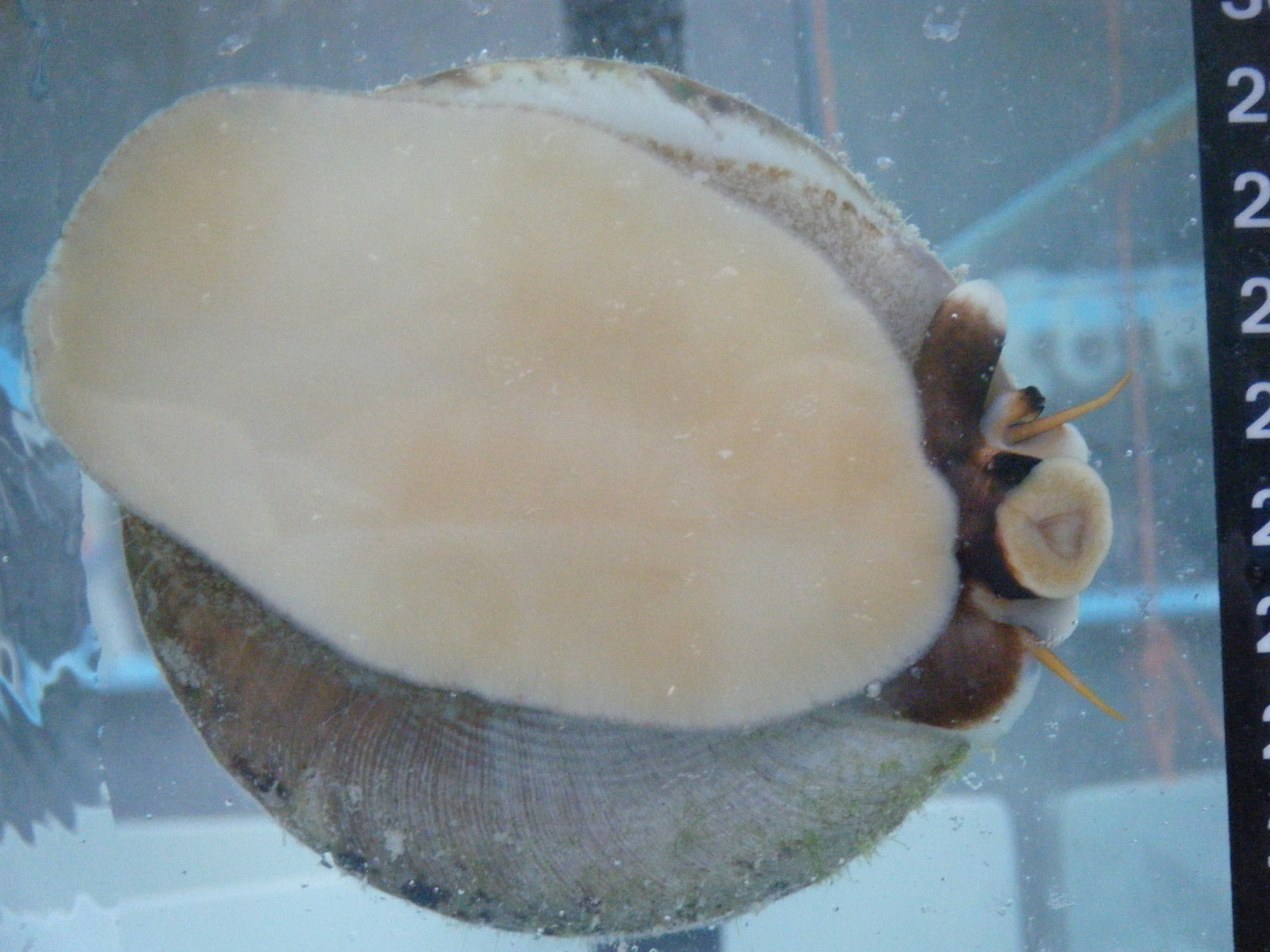
The animal inside the shell is mostly made up of a large muscular foot (Ruppert, Fox and Barnes, 2004), which is brownish orange on the topside and creamy white underneath. This foot can be seen in the photo to the right, as the large creamy white structure adhering to the side of the tank and taking up most of the photograph.
Along from the foot the head is visible. It is circular and creamy white. The tongue like radula can be seen within the mouth, which the animal uses when feeding (Ruppert, Fox and Barnes, 2004).
Two orange cephalic tentacles protrude either side of the head (Ruppert, Fox and Barnes, 2004). These are clearly visible in the photo, and contain tactile and chemo-receptor cells used for sensory functions.The eyes are located at the base of these cephalic tentacles. The eyes are not well developed, containing only photoreceptor and pigment cells (Nash, 1985). This means they only serve in light detecting functions, rather than image forming.
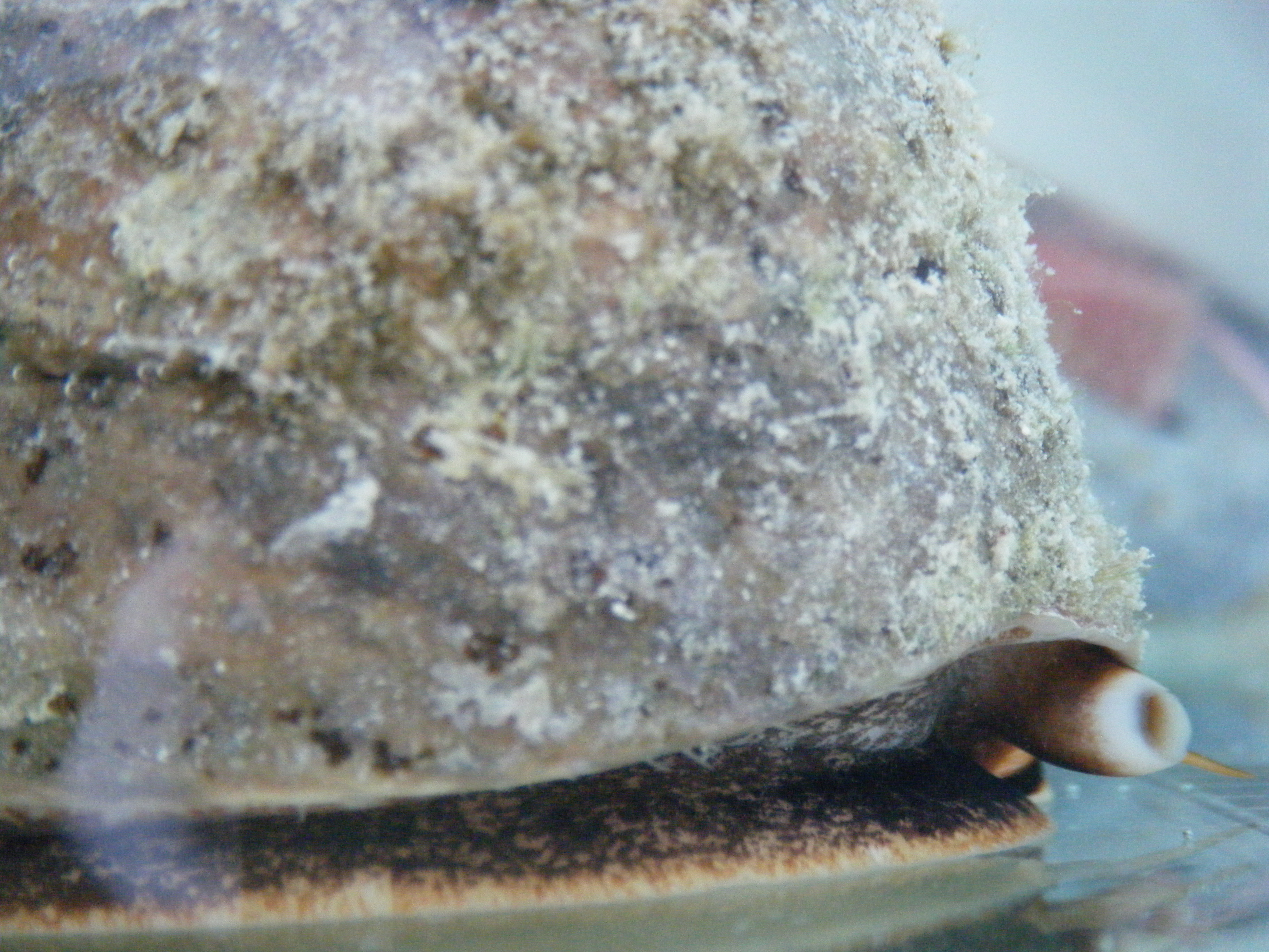
 To the left of the head, when looking at the photo on the left, a brown tube- like opening is visible. This is the animal’s anus, and is located close to the mouth due to torsion during larval development (Ruppert, Fox and Barnes, 2004). It is clearly visible protruding from the side of the animal in the photo below. To the left of the head, when looking at the photo on the left, a brown tube- like opening is visible. This is the animal’s anus, and is located close to the mouth due to torsion during larval development (Ruppert, Fox and Barnes, 2004). It is clearly visible protruding from the side of the animal in the photo below.
The gills and osphradia are located behind the head, just under the shell (FA0, 1999). Gills are used for respiration while the function of osphradia is uncertain. Most scientists tend to believe they are used to detect sediments passing over gills, so they do not get caught in the gills (Ruppert, Fox and Barnes, 2004).
Just behind the anus is the gonaductus opening, which is where the gonads are released during spawning (FAO, 1999).
According to Asano (1963) there are two phenotypic variants of this species. The first has a shell that is conical, with straight sides and a flat base. In the second the final shell whorl expands greatly to form a wide base.
|