Internal Anatomy
Figure 1: Cryptoplax larvaeformis dissection, detailed are the main components of the digestive and excretory systems.
The beginning:
The mouth is found on the ventral side on the Chiton, and in the center of it's head. The mouth passes into the buccal cavity (or Gullet), which is highly muscularised and strengthened by chitine, forming a firm under layer for the radula (Kaas & Van Belle 1893).
The radula:
The Chiton radula is highly conserved over the entire class, and differing from some other molluscs is quite simple. It can extend as far back as the 3rd or 4th shell plate, and is located ventrally (bellow) of the digestive tract. By means of a complicated muscular system the radula is pushed into the pharynx of the Chiton and is moved back and forth to scrape it's food off the substratum (Dell’Angelo & Smriglio 2001). Cryptoplax larvaeformis is a herbivorous Chiton and therefore it's radula is designed for scrapping algae of rocks and other substrate (Littler & Littler 1999).

Figure 2: Internal look at the radula of a cryptoplax lavifmious. A. The entire radula, demonstrating it's location ventral to the digestive tract. B. Highlights the musculature involved in the protrusion of the radula. C. Here the muscles involved in the movement of the radula are again highlighted, as well as an unknown structure, a possible extension of the hemal system, highly integrated with the radula.
The Stomach:
The food is moved by the radula into the buccal cavity, mucus supplied by a pair of salivary glands mixes with the food moving it into the esophagus. Once in the stomach, digestion involves large glands and enzymes produced by the digestive cecum.
The end:
The Intestine:
The intestines are about 4 or 5 times the length of the chiton and are divided into the anterior part of the intestine and the posterior part. The fecal pellets are formed in the posterior part of the intestine and are ejected through the anus which is situated behind the foot, and usually placed on a papilla (Kaas & Van Belle 1893).
Figure 3: Microscopic view of a cryptoplax lavifmious intestine. A. An extracted intestine. B. The anterior and posterior parts in the intestine. C. Anterior part of the intestine demonstrating the formation of the fecal pellets.
Excretion:

The excretory system involves a pair of large nephridia (Figure 4) located ventrally on either side of the visceral cavity. Residual products in the hemal system are delivered to the nephridia by pores and renopericardial canals in which are highly cilliated. excretory products are expelled via a tube that delivers these products to the nephridiopore which is located in the pallial groove among the most posterior gills (Kaas & Van Belle 1893, Dell’Angelo & Smriglio 2001) (figure).
Nervous System:
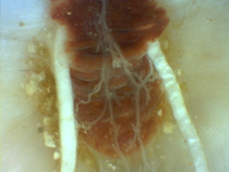
The Chiton nervous system consists a structure called the buccal nerve ring and buccal ganglia, these structures perform similar functions as a brain would. From here extends the paired longitudinal lateral nerve cords and the paired pedal or ventral nerve cords. The longitudinal lateral nerve cord is often connected at the posterior end to form a large loop around the entire body. Nerve cells innervated by the lateral cords penetrate the shell plates and ascend vertically through pores in the layers of the plate. These pores and complex nerve cells allow for the use of aesthetes embedded in the shell. (Kaas & Van Belle 1893)

Figure 5: Microscopic picture of nerves in cryptoplax lavifmious overlaying the main artery
of the hemal system.
|