Overview
Brief Summary
Physical Description
Size and Appearance
Ecology
Distribution and Habitats
Microhabitats
Interactions
Life History & Behaviour
General Behaviour
Feeding and Predation
Reproduction and Life Cycle
Evolution & Systematics
Phylogenetics
Morphology and Physiology
External Morphology
Internal Anatomy
Cell Biology
Genetics
Nucleotide Sequences
Conservation
Threats and Conservation
Wikipedia
References & More Information
Bibliographies
Biodiversity Heritage Library
Search the Web
Names & Taxonomy
Related Names | External Morphology
Although Stichopus chloronotus is called the Greenfish and is dark green in colour (Baker 1929), it frequently appears to be black. It is quadrangular in transverse section and has papillae along the angles of its shape (Baker 1929). The integument is thick and smooth, and the dermis is made up of multiple layers including a superficial zone, a middle zone of collagen fibres, and a hypodermis (Menton and Eisen 1970). Although they generally feel soft, the body wall can harden and make the animal quite stiff (Motokawa 1982). This is achieved through modifications in the chemical properties of the mutable collagenous tissue, which are then reflected in the mechanical properties of the dermis (Motokawa 1982). The stiffening and softening agents used in this process are found in the coelomic fluid and are activated when the dermis is stimulated (Motokawa 1982). Tube feet are found on the dorsal surface of the body and are used for walking. The feeding tentacles are leaf shaped, as is characteristic of the Stichopodidae family (Edgar 2008). The feeding tentacles are actually modified tube feet (Edgar 2008), and there are 20 present around the mouth (Conand et al. 2008). The average length of the feeding tentacles is approximately 5 mm (Conand et al. 1998). These feeding tentacles are used to collect sediment and bring it to the mouth (Conand et al. 1998).
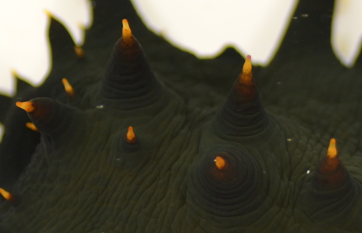
Image 1: Shown here are the orange-tipped papillae of Stichopus chloronotus.
|
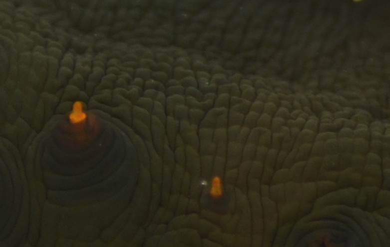
Image 2: The papillae and wrinkled-looking dermis of S. chloronotus.
|

Image 3: The tube feet of S. chloronotus. They are able to stretch, break off, and be regenerated. |
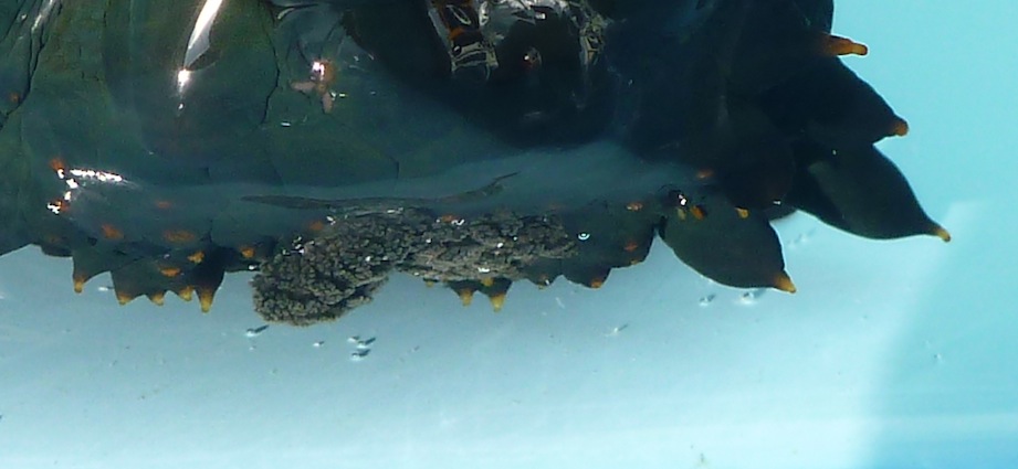
Image 4: The feeding tentacles of S. chloronotus. Although they are extended when feeding, generally these tentacles are gathered up so that they are not seen. |
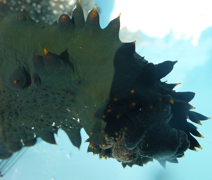
Image 5: The anterior (head) end of Stichopus chloronotus. Visible are the papillae, tube feet, and feeding tentacles. |
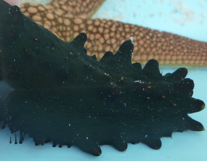
Image 6: The tube feet of Stichopus chloronotus can be used to hold onto hard surfaces, as shown in this image. They can also break off if stretched too far (bottom right of image), but they are later regenerated. |
|
|