INTERNAL ANATOMY
Like other echinoderms, H. edulis has a water vascular system (WVS), which is a multi-purpose system composed of canals that connect the tubed feet. The WVS is involved in locomotion, feeding, gas exchange and other tasks, and works by controlling water flow through the canals, similar to a hydraulic system [14].
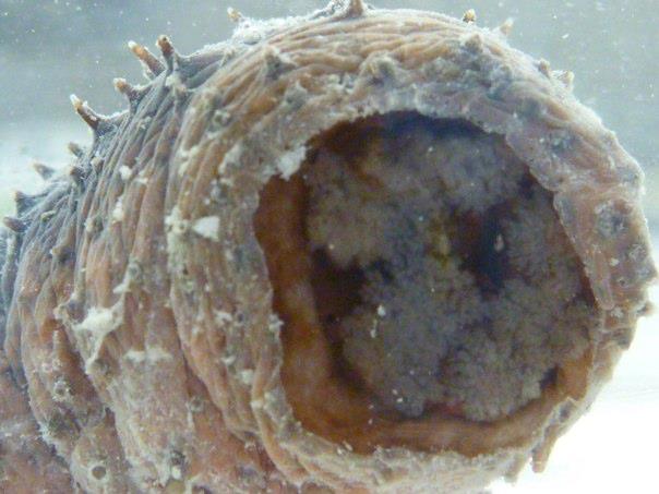
Inside the mouth H. edulis has buccal podia (tentacles), which are specialised oral tube feet. These are connected to an introvert, which is extended and retraced in conjunction with the buccal podia [14]. When they are exposed, they are primarily are used for feeding. Otherwise they are retracted into the buccal cavity. Buccal podia are also associated with inverting behaviours and use this, in conjunction with tube feet, to invert themselves back to an upright position if they have been flipped.
Buccal podia of H. edulis
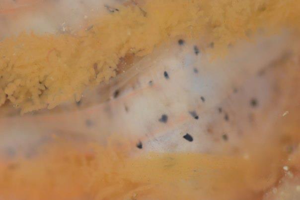
Although the buccal podia and tube feet function as gills, the chief gas exchange organ in H. edulis occurs in the form of a bilateral pair of respiratory trees or water lungs. Each tree arises as a diverticulum from the wall of the cloaca and branches repeatedly to form a system of hollow, blind-ended tubes [14]. Respiratory trees are ventilated through muscular pumping of the cloaca and through contraction of the respiratory trees themselves.
Respiratory Trees of H. edulis
Although many holothurians have cuvarian tubules, which are used as a defence mechanism, dissections of H. edulis suggest this species lacks such defence mechanisms and may instead rely on chemical defences.
Holothurians have long tubular gut. This is an adaptive requirement to deal with the poor nutrient quality of their diet. The long gut allows for maximum absorption to occur.
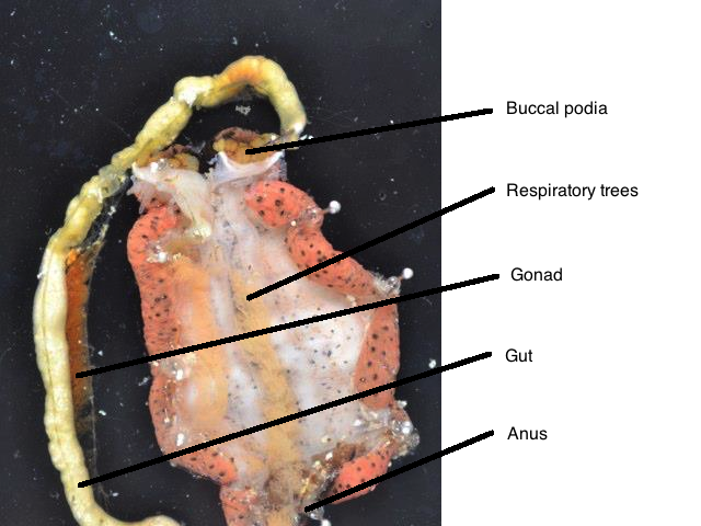
Internal anatomy of H. edulis |