Internal Anatomy
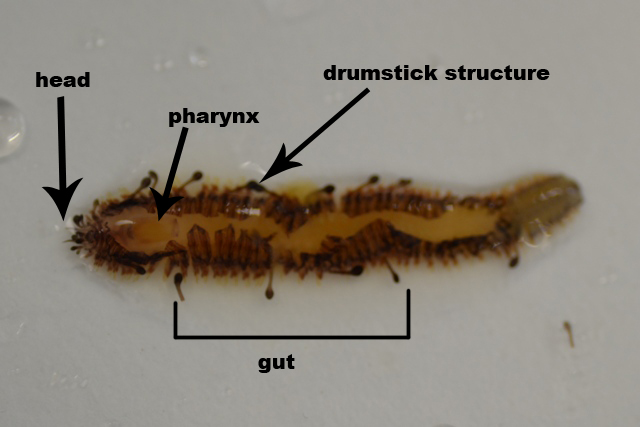 |
an overview of the internal organs of g. clavigera
|
Epidermis
The epidermis in polychaetes is composed of a cellular monolayer that is covered by a cuticle. The epidermis inhabits secretory cells, sensory cells and as its main component supportive cells (Hausen, 2005).
Supportive cells
Supportive cells show different cell morphologies, it reflects different physiological specializations.
Secretory cells
They are abundant in the polychaete epidermis and are situated separately between the supportive cells or maybe grouped within glandular fields or form complex multicellular glands
Coelomic cavity
There is no evidence showing that g. clavigera has a coelomic cavity, thus it is considered as acoelomic.
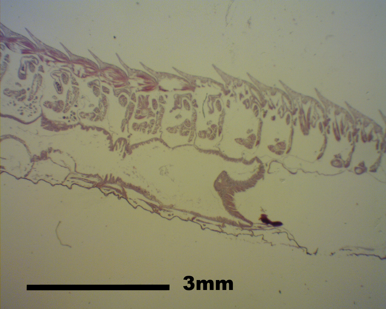 |
A cross section of g.clavigera body
|
Muscular system
Polychaetes are soft-bodied animals, their mobility and the maintenance of the body are affected by their muscular system (Tzetlin & Filippova, 2005). The polychaete muscular system consists of several components, such as circular and longitudinal fibres of the body wall, parapodial, chaetal, oblique, diagonal, dorsoventral fibres, as well as muscular structures elements of septa and mesenteries.
circular muscles
circular and other transverse fibres are usually found below the extracellular matrix of the integument and are poorly developed compared to the longitudinal muscles underlying the layer of the circular muscles if present (Westheide & Rieger, 1996).
longitudinal muscles
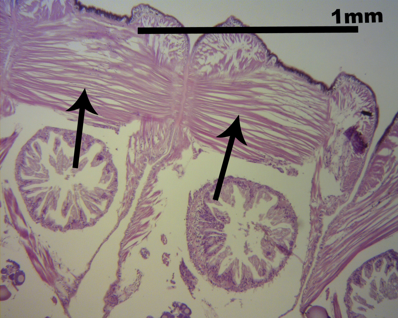
Longitudinal muscles run along the whole body length and usually form discrete bands. The fibres making up the bands are arranged in different patterns. A band maybe formed by large flattened cells lying in a single row and not being covered by the coelomic epithelium. The nuclei of these cells are usually located on the distal part facing the coelomic cavity (Tzetlin & Filippova, 2005).
|
longitudinal muscles (black arrows) in the polychaete
|
Parapodium muscle complex
The data on the muscle arrangement in parapodia are limited.
Excretory system
According to Bartolomaeus (1999), the family polynoidae (g. clavigera belongs to the family polynoidae) involves protonephridia and metanephridia for filtration.
Digestive system
The alimentary canal of polychaetes consists of three parts: foregut, midgut and hindgut. The structure of the gut is correlated with adaptations to feeding and life style of polychaetes (Purschke & Tzetlin, 2005).
Foregut
It is originated from ectodermal, it is formed by stomatodeal and proctodeal invaginations of the ectoderm and thus usually bearing a cuticle. It gives rise to the buccal activity, pharynx and oesophagus.
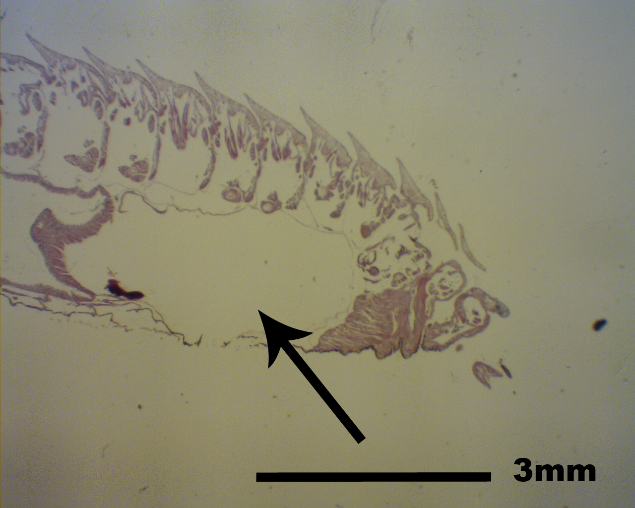 |
the foregut (black arrow)
|
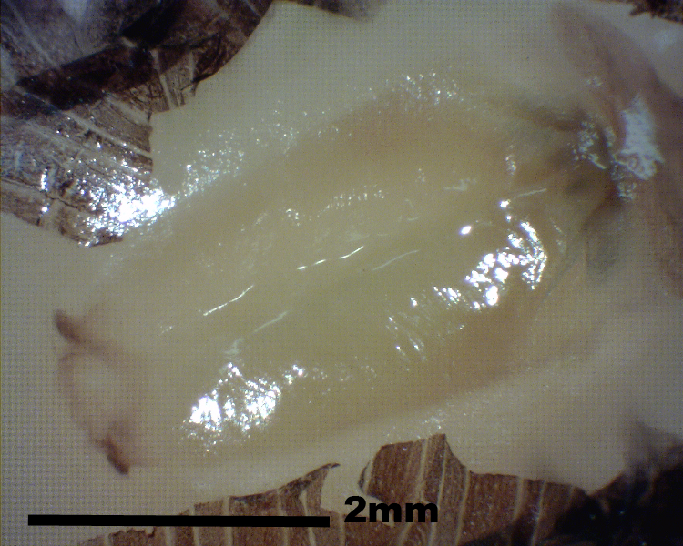 |
The pharynx of g. clavigera
|
Midgut
It is derived from the endoderm. It can be divided into a stomach and the intestine proper.
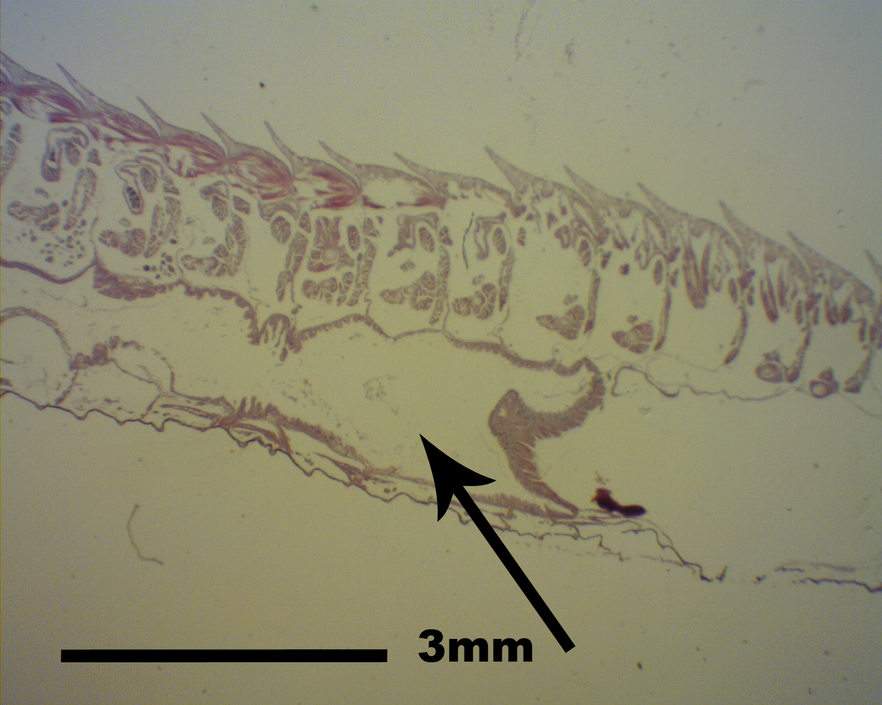 |
The foregut of the polychaete (black arrow)
|
Hindgut
It is also originated from ectodermal. It normally forms only one part.
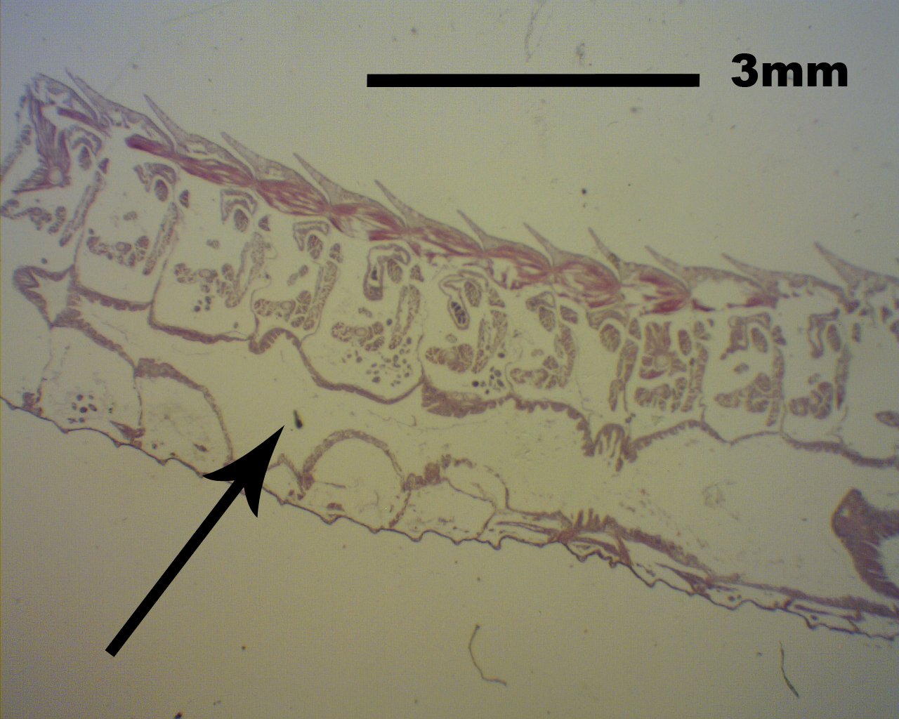 |
The hindgut of the polychaete (black arrow)
|
|