Overview
Brief Summary
Physical Description
Size
Identification Resources
Ecology
Local Distribution and Habitats
Biogeographical Distribution
Micro-habitats and Associations
Crypsis
Life History & Behaviour
Behaviour
Reproduction
Settlement Induction
Evolution & Systematics
Fossil History
Systematics or Phylogenetics
Morphology and Physiology
External Morphology
Internal Anatomy
Molecular Biology & Genetics
Nucleotide Sequences
Molecular Biology
Wikipedia
References
Bibliographies
Names & Taxonomy
Synonyms
Common Names | Reproduction
|
Fertilisation

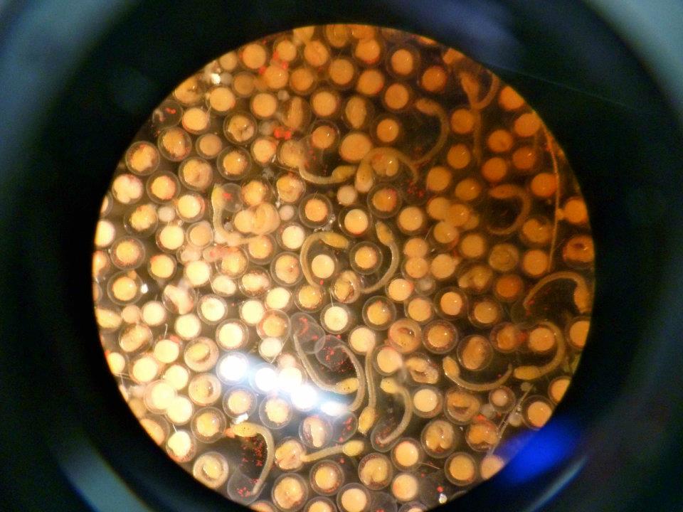
Figure 1. In vitro fertilisation experiment
|
Although individual red-throated ascidians contain both male and female sex cells, i.e. are hermaphrodites, they are self-sterile and unable to fertilise their own eggs. Eggs and sperm are expelled from individuals into surrounding seawater in the hope that they will meet with gametes of the same species. In order for eggs to stay afloat, follicle cells balloon, creating buoyancy and increasing the chance of fertilisation. Spawning events occur throughout the year in most tropical populations yet are typically seasonal in sub-tropical populations (Degnan 1991, Shenkar & Loya 2008).
|
|
Development
After fertilisation has occurred, development of H. momus is typical of other ascidians and relatively rapid. Larvae hatch between 10 and 20 hours after fertilisation at temperatures between 21 and 28 degrees celsius respectively (Degnan et al. 1996). Shortly after fertilisation, cytoplasmic rearrangement results in an orange endoplasm, white myoplasm and grey-green ectoplasm, each of which is inherited by the blastomeres (figure 2A – D). When held at 25 degrees, 40 minutes marks the first cleavage (figure 2A) which is then followed by another cleavage every 15 minutes for the next hour. The number of cells doubles with each cleavage, resulting in 64 cells at about 2 hours after fertilisation (figure 2D). After this time, cell division slows and is no longer synchronous as cell specialisations begin to occur during a process called gastrulation. Here, cells located at the vegetal pole (yolk end) move towards the inside of the embryo to create an indent called the gastrula; this will later become the anus. After 4 hours has passed, the beginnings of a brain form over the vegetal hemisphere and animal cells expand and cover the entire embryo. At 5 hours, the embryo starts to become elongate along that anterior-posterior axis (Fig 2E). By 6 hours, the notochord cells begin to form the beginnings of a tail (Fig 2F). Shortly after 7 hours, the notochord and tail becomes longer and the spinal cord appears (Fig 2F– H). At 9 hours, eye pigmentation occurs and the larval test begins to form. After just 10 short hours, the tadpole larvae will hatch (Fig 2I).
|

Figure 2. Developmental stages of Herdmania momus larvae (see above text for description) |
|
Metamorphosis
After larvae hatch they require at least an additional 3 hours of development before they become competent to settle and metamorphose (Degnan et al. 1996). Resorption of the tail is one of the first morphological signs that metamorphosis has begun and is accompanied by formation of the adult tunic and test cells outside the epidermis (figure 3A-C). Most larvae then attach to the substratum on their side, rather than their adhesive papillae (figure 3A). Once attached, the body rotates so that the attachment organs project into the tunic and the adult body axis is established. The larval muscles then degenerate, the endostyle, pharynx and gut loop become discernible. After 5.5 days from induction, the atrial and branchial siphons extend through the tunic and muscle cells form around them. After about a week the juvenile is fully formed with all adult tissues visible, apart from the gonads.
|

Figure 3. Metamorphosis stages of Herdmania momus
|
|
|