Overview
Summary
Physical Description
Size
Colour Identification
Ecology
Local Distribution and Habitats
Biogeographical Distribution
Life History & Behaviour
Behaviour
Reproduction
Evolution
Chemical Evolution
Systematics or Phylogenetics
Morphology and Physiology
External Morphology
Internal Anatomy
Histology
Molecular Biology & Genetics
Nucleotide Sequences
Conservation
Threats
References & More Information
References
Search the Web
Names & Taxonomy
Synonyms & Common name
Acknowledgements
Acknowledgements | Histology
A T.bayeri from Shark Bay, Heron Island was brought back to The University of Queensland for sectioning and staining. Histology information is based on Jensen 1992; Wagele 2000; Wagele 2005; Wagele & Johnson 2001; Wagele et al. 2010. The specimen was cut longitudinally. However, it would be clearly to see if the specimens was a transverse cut cross sectionally.
Figure 1: T.bayeri sectioned under low power microscope.
Figure 1 show the whole specimen histology. The anus should be on the left of the pictures. However, because the specimen curl up during fix, the specimen is poorly sections. The pictures also indicate that there are a lot of special granular along the body.
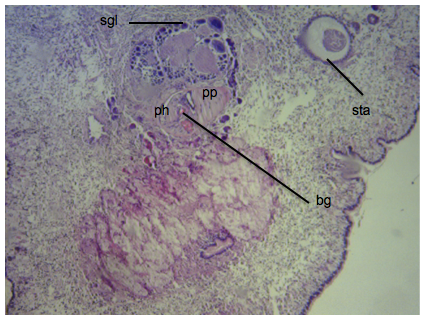
Figure 2: Sections near the head of T.bayeri
Abbreviation: Buccal gangion (bg), Pharynx (ph), Pharyngeal pouch (pp), Salivary glands (sgl), statocyst (sta)
Figure 2 emphasized the head region of T.bayeri where pharynx can clearly observed. Moreover, statocyst is also present. Statocyst help organisms to maintained the equilibrium with respect to gravity, which help them when clawing on the vertical surface. It is possible that the mass below the pharynx is unknown and no literatures account for about it yet.
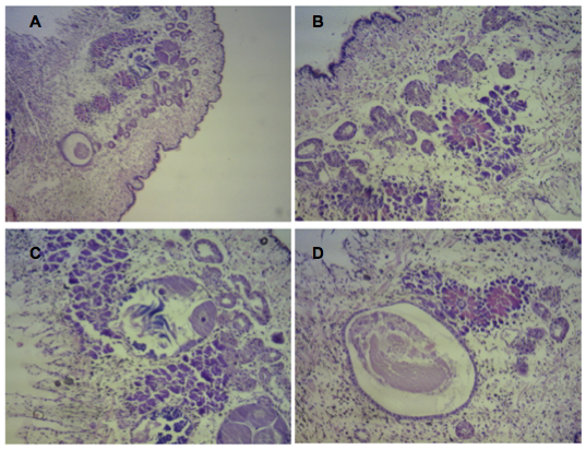 Figure 3: Histology of Thuridilla bayeri.
Figure 3 showing various part of the sections. A) showing a big pictures of the sections, again statocysts can be seen clearly. B) showing flower-like structure, which suspect to be a gland surrounded by tubules. This structures can be seen from the anterior end of the body through to posterior end. C) showing that look like gonads, however, it is still unknown. D) showing big mass which located just at the end of the head region. With it location, it is possible that it's a mucus glands.
Information on the histology of Thuridilla bayeri is limited. There were no literatures found focusing on the histology of this species. To analyze this images, information on histology of Sacoglossa was applied with the help of position of internal anatomy. Further studies should be done to look through the histology of this species, as many structures look completely different from typical Sacoglossan. Understanding their unique histology may allow us to understand the evolution and phylogeny of the species better.
|
|